An optimal criteria for identification of Trichomonas spp. by digital image from iQ200TM SPRINT urine analyzer
Author'(s): Wuttichai Suksanit1*, Yuwin Aniwataugkoorn2, Yotsombat Changtrakul1 and Kutcharin Phunikhom1,3
1Department of Diagnostic Microscopy Unit, Srinagarind Hospital, Faculty of Medicine, Khon Kaen University, Thailand.
2Faculty of Medical Technology, Khon Kaen University, Thailand.
3Department of Pharmacology, Faculty of Medicine, Khon Kaen University, Thailand.
*Correspondence:
Wuttichai Suksanit, Department of Diagnostic Microscopy Unit,Srinagarind Hospital, Faculty of Medicine, Khon Kaen University,Thailand.
Received: 20 February 2020; Accepted: 07 March 2020
Citation: Wuttichai Suksanit, Yuwin Aniwataugkoorn, Yotsombat Changtrakul, et al. An optimal criteria for identification of Trichomonas spp. by digital image from iQ200TM SPRINT urine analyzer. Cancer Sci Res. 2020; 3(1); 1-3.
Keywords
Introduction
The routine urine analysis services are important in helping diagnose and assess kidney function initially, also an indicator of whether there is a pathological condition in the kidneys. Especially disorders related to the urinary system.
The detection and reporting of urinary sediment can help indicate the clinical relationship of the disease. As well as the detection and reporting of the Trichomonas species (spp.). This can be transmitted via sexual transmission. And most people who are infected about 70% do not show symptoms but can be transmitted and can cause the disease, such as vaginitis in women, the cause of irritation due to urethritis in men. Therefore, the detection or report of Trichomonas spp. is clinically important to help physicians diagnose patients correctly.
Nowadays, routine examination of urine analysis depends on physical examination, chemical examination and microscopic examination. Microscopic examination to separate various types of urine sediment. It is important for reporting results to help doctors diagnose patient results correctly, precise and efficient.
At present the unit has introduced the iQ200TM SPRINT urine analyzer that uses digital imaging principles for routine. The reason was the users of the iQ200TM SPRINT urine analyzer do not have criteria and methods to review and notice that the device can separate the Trichomonas spp. parasites from leukocytes or other urine of similar size. May cause reports of Trichomonas spp. inaccuracy and error. This study interested to find various observations from the images obtained from the principles of the machine that can detect or report the Trichomonas spp. by the iQ200TM SPRINT urine analyzer to get accurate results. It is beneficial to with accuracy and maximum efficiency.
Objectives
- To study an optimal criteria for identification of Trichomonas by observe from the images obtained the principle of the iQ200TM SPRINT urine analyzer.
- To isolate the Trichomonas from white blood cells or other types of urine of similar size.
Materials and Methods
- Collecting and studying urine analysis samples at Srinagarind Hospital Faculty of Medicine Khon Kaen University by images from the iQ200TM SPRINT, it is suspected to be a Trichomonas spp., parasite such as blurry images, unclear images in various forms. And / or other characteristics other than cells that are directly classified by the device, 22 cases (N=22).
- Take the urine sample that has images as in to examine with a microscope to compare and confirm the images from the iQ200TM SPRINT that is Trichomonas spp. Based on optimal criteria the suspected images for trich in “WBC” and “UNCL” categories were analyzed.
- Recording various characteristics of the suspected images and Trichomonas spp.
- Collected data obtained for analysis and conclusion.
Results
From the criteria for Trichomonas spp. investigation by various types of photos obtained from the iQ200TM SPRINT urine analyzer. N = 22 were 18 female and 4 males. There are 2 females and 2 males, for a total of 4 reported by the WBC or cells detected in UNCL, were suspicioused for Trichomonas spp.. Then examined by a microscope has a negative result that is not Trichomonas spp. (false positive found 18.18% (4/22)).
Therefore, the following observations:
- Flag or note by machines are WBC,
- Cellular photo features: Cytoplasm resembling a flagella or pseudopod as shown in figure
- The cell images is unclear, as shown in figure
- The combination with leukocytes esterase, there is a positive result from trace to 3+.
By characteristics or observations according to figures 1 and 2, confirmed by microscopic examination, it was found that negative results were not Trichomonas spp.
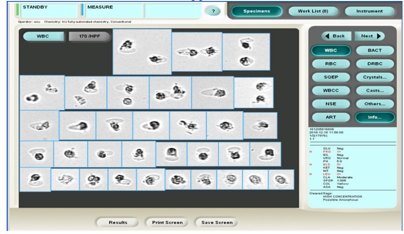
Figure 1: The characteristic of white blood cells (WBC) suspected of being Trichomonas spp. (false positive).
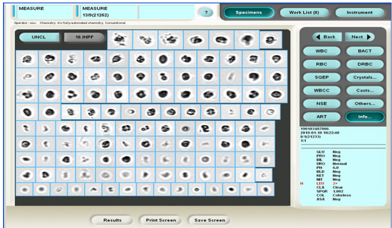
Figure 2: The various cells with blurred images in UNCL and suspected to be a Trichomonas spp. (false positive).
In most cases, the WBC report is found from 5-10 cells / HPF to more than 100 cells / HPF. As an example shown in figures 1 and 2, and summarized in table 1.
Table 1: Image data from the WBC iQ200TM SPRINT, Luekocyte esterasce and the detected amount but not the Trichomonas spp.
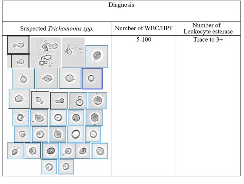
A total of 18 patients gave positive results when confirmed with a microscope Trichomonas spp. was 81.82%, which can conclude that an optimal criteria for identification of Trichomonas spp. as follows:
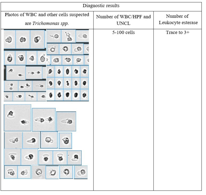
- Flag devices are WBC and UNCL, there is a smooth cell (homogeneous) similar size may be small or older than WBC, with all WBC detected and leukocytes esterase positive results from trace to 3+.
- Cells with flagella or pear shaped may be found (as in figure 4-6).
- Confirmation of the Trichomonas by microscopy
In this study, positive results (Trichomonas spp.) leukocytes and leukocyte esterase were all positive. In which white blood cells were found from 5-10 cells / HPF up to more than 100 cells / HPF and leukocyte esterase have a positive result from Trace to 3+, as shown in figure 3 to 6. And show the summary in table 2.
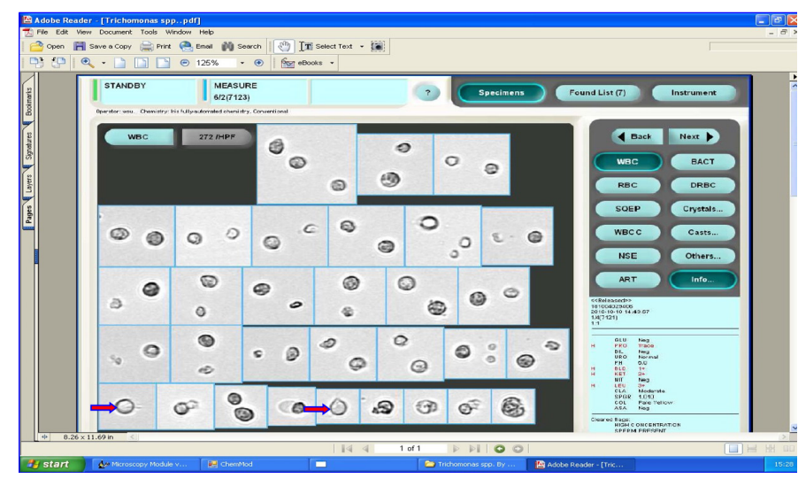
Figure 3: The nature of the cell suspected to be the Trichomonas spp.found in the WBC channel (arrow pointing).
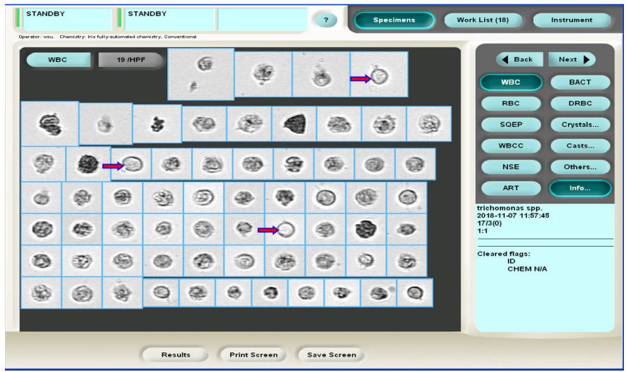
Figure 4: The characteristics of Trichomonas spp. found in the WBC channel and report as Trichomonas spp. (arrow pointing).
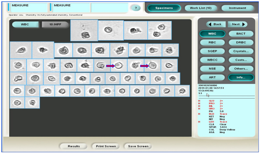
Figure 5: The nature of the cell suspected to be the Trichomonas spp. found in the WBC channel (arrow pointing).
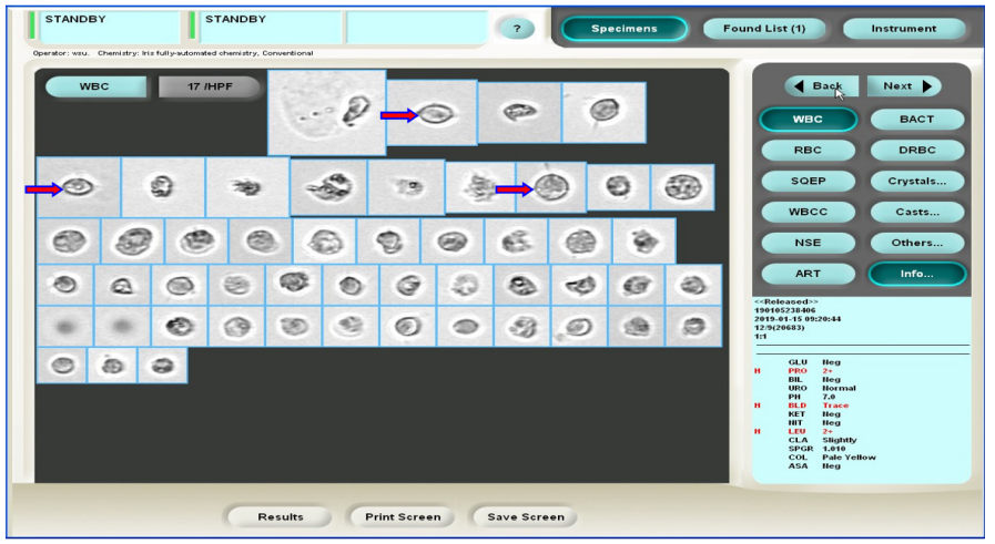
Figure 6: The nature of the cell suspected to be the Trichomonas spp. found in the WBC channel (arrow pointing).
According to the results of the 22 automatic sediment screening tests using the iQ200TM SPRINT. Methods for viewing and observing the characteristics of the above images including the principles that the image classification system (Categories) of all urine sediments it was found that the device was able to correctly classify WBC images. It may look like some images that are unclear and suspect that it may be a Trichomonas spp. in figure 1, 2. A total of 4 cases wanted to do a negative control. The confirmation by microscopy it was not Trichomonas spp.
Table2: Image data from the iQ200TM SPRINT that uses the Trichomonas spp. WBC amount, Luekocyte esterase and the detected amount.
Discussion and Conclusion
In summary, the images of the Trichomonas spp. found in this study are shown in figures 3 to 6 and table 2. After confirm with microscopic examination, it was found that all Trichomonas spp. (positive 81.82%). Suspected images of Trichomonas spp. negative findings were the active WBC with pseudopodia or WBC with bacteria whereas the true Tricomonas spp.were always more hematogenously seen with flagella. The presence of WBC in the absence of bacteria or yeast is the clue to look for the pear shape body and flagella. The cross-check generated by iQ200TM series analyzers, there flag for trichomoniasis, and it also useful for Trichomonas spp. identification. Our data suggest that utilization of this optimal criteria contribute to increase the sensitivity in the diagnosis of trichomoniasis. However, the varieties of suspected Trichomonas spp. images must be considered. The continued viability of parasites and well-trained microscopist are also required for correct diagnosis.
References
- Kirby H. The structure of the common intestinal tichomonad of J Parasital.1945; 31: 163-175.
- Manual of the urine analyzer iQ200 SPRINT Diagnostic Microscopy Unit Srinagarind Hospital Faculty of Medicine Khon Kaen
- iQ200- Image Encyclopedia. 300-6160-RevB.pdf
- Beckman iRICELL3000. 2016. Available from: URL: https://www.beckmancoulter.com/wsrportal/wsr/diagnostics/ clinical-products/urinalysis/iricell-workcells/iricell3000/ index.htm