Virtual Sonographic Folliculoscopy for Monitoring of Ovarian Stimulation in "Poor Responder" Women Prepared for in vitro Fertilization
Author'(s): Tesarik Jan1*, Mendoza-Tesarik Raquel1 and Mendoza Nicolas1,2
1MARGen Clinic, Granada, Spain.
2Department of Obstetrics and Gynecology, University of Granada, Granada, Spain.
*Correspondence:
Jan Tesarik, MD, PhD, Director, MARGen Clinic, Camino de Ronda 2, 18006 Granada, Spain, Tel: +34 606376992; E-mail: jtesarik@clinicamargen.com.
Received: 19 September 2017 Accepted: 27 October 2017
Citation: Tesarik Jan, Mendoza-Tesarik Raquel, Mendoza Nicolas. Virtual Sonographic Folliculoscopy for Monitoring of Ovarian Stimulation in “Poor Responder” Women Prepared for in vitro Fertilization. Gynecol Reprod Health. 2017; 1(3): 1-3.
Abstract
Rationale: It is known that oocytes fail to be recovered from some large follicles observed in human ovaries by conventional ultrasound examination. The prediction of oocyte recovery is particularly important when planning in vitro fertilization in women with low follicle numbers.
Objective: To describe a new ultrasound-based imaging technique, to which we have given the name virtual sonographic folliculoscopy, and to evaluate its ability to predict oocyte recovery from patients with poor response to ovarian stimulation.
Methods: We report a technique for visualization of the cumulus oophorus within the cavity of antral ovarian follicles, based on transformation of 2-dimensional sonographic recordings, taken from the vagina, to 3-dimensional virtual reconstructions. The prediction of the presence of an oocyte-cumulus complex within the follicles is confronted with the actual yield of oocytes by follicle aspiration.
Results: The cumulus-oocyte complexes were clearly visible within some, but not all, large antral follicles. In a group of 20 women with poor response to ovarian stimulation, treated by in vitro fertilization, the presence of a cumulus-oocyte complex in aspirated follicular fluid confirmed the folliculoscopy-based prediction in 17 cases.
Conclusions: Virtual sonographic folliculoscopy appears to be a useful tool for the prediction of oocyte recovery from antral follicles resulting from ovarian stimulation of poor responder patients.
Keywords
Introduction
Various techniques for the examination of female pelvic organs are available nowadays, but only few can generate 3-dimensional images of organ cavities. Some of them, such as computerized tomography and magnetic resonance imaging, are able to generate images whose precision and spatial resolution is equal, or even superior, to that of the conventional endoscopic techniques [1].
However, these techniques require patient exposure to factors, such as ionizing radiation or iodinated contrast media, which might be harmful to the reproductive organs. Thus, there is growing interest in employing techniques based on ultrasound in the pelvic region [2,3].
We have recenly reported a case of an intrauterine synechia, detected by ultrasound-based virtual hysteroscopy, and subsequently confirmed and removed by conventional operative hysteroscopy [4]. Here we show how the aplication field of this technique can be extended to antral ovarian follicles, cavities that are not accessible to conventional endoscopic techniques, and how the information obtained can be used in taking important therapeutic decisions in protocols of ovarian stimulation in poor responder women, in whom only few antral follicles develop.
Methods
The technique of virtual sonographic folliculoscopy consists of two sequential phases: image acquisition and image processing. The former phase is a direct examination session with the patient and takes a relatively short time. The latter, usually more time- consuming, is done in the absence of the patient.
During the phase of image acquisition a short sequence of 2-dimensional images of antral ovarian follicles was recorded from the vagina with the use of 11CV3 transvaginal probe (Toshiba Medical Systems Europe, Zoetermeer, The Netherlands). The planar 2-dimensional recordings, taken during this first phase of examination, were subsequently converted into 3-dimensional virtual reconstructions of the antral cavities examined, with the use of the “fly-through” technology (Fly Thru, Toshiba) incorporated in the ultrasound machine (Aplio 500 Platinum). During this phase, the operator can “fly” freely through the virtual antral cavity and select different angles of view. The direction of movement through the cavity examined was varied in each follicle examined, in search for the cumulus ophorus.
This study involves 20 patients with low ovarian reserve, as defined by the Bologna criteria [5], undergoing ovarian stimulation for an in vitro fertilization (IVF) attempt. Only follicles with the mean diameter comprised between 10 and 24 mm were evaluated. This evaluation was performed on the day of ovulation induction by the injection of hCG.
Results
Virtual folliculoscopy was used in the context of ovarian stimulation of poor-responder patients who developed only few antral follicles, with the aim to identify the cumulus oophorus in order to estímate the number of oocytes which could be eventually recovered by follicle aspiration. The cumulus oophorus was occasionally seen in the initial 2-dimensional recordings of antral follicles. However, in most cases, its clear identification was only posible in the reconstructed 3-dimensional virtual images (Figure 1).
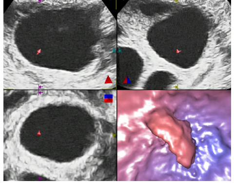
Figure 1: Three complemetary 2-dimensional recordings (both upper panels and left lower panel) used for virtual reconstruction and the resulting 3-dimensional image (right lower panel) of an antral follicle cavity showing a cumulus oophorus mass.
It was hypothesized that the detection of the cumulus oophorus by virtual folliculoscopy could predict successful oocyte recovery by subsequent follicle aspiration. Accordingly, the expected number of oocytes retrieved by folicular aspiration in each patient was defined as the number of antral follicles with a cumulus oophorus, detected in the patient’s ovaries on the day of ovulation induction. The comparison of the expected and the real number of oocytes recovered from each patient is shown in Table 1. From the 20 patients involved in this study, both numbers were identical in 17 of them. The real number of oocytes was lower than the expected one in 2 cases and higher in 1 case (Table 1). In one case, in which no oocyte was recovered, this result was accurately predicted by virtual folliculoscopy, but the couple wished ovarian puncture to be performed in spite of this adverse prediction.
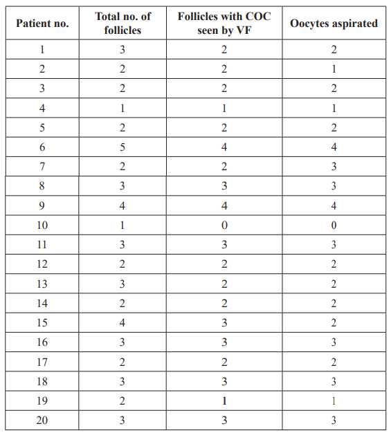
Table 1: Comparison of the number of oocytes predicted by virtual folliculoscopy (VF) and the real number of oocytes actually recovered by follicular aspiration.
Abbreviations: COC, cumulus-oocyte complex; VF, virtual folliculoscopy.
Discussion
Virtual sonographic folliculoscopy is a new application of the Fly Thru technology in gynaecology. The early attempts at visualizing the cumulus oophorus within antral follicles in the context of ovarian stimulation date from the 1980s [6]. Even though the detection of the cumulus by 2-dimensional ultrasonography is questionable, the introduction of 3-dimensional volume scanning techniques made the visualization of the cumulus mass more performant [7]. The use of virtual folliculoscopy, as described in this study, provides further refinement of sonographic detection of the cumulus oophorus, which may be particularly important for taking the right decision and enabling adequate patient counselling in cases of poor ovarian response with only few growing follicle- like structures some of which might actually be empty follicles or cysts.
In fact, the number of oocytes recovered by follicle aspiration can be predicted by this technique with a relatively high accuracy. Out of 20 patients examined, the predicted and real numbers of oocytes recovered was concordant in 17 cases. In 2 of the 3 discordant cases, an oocyte failed to be recovered from 1 follicle in which the cumulus was detected by virtual folliculoscopy, possibly because of the retention of the cumulus-oocyte complex during follicle aspiration. In one case, unexpectedly, 3 oocytes were recovered although the cumulus oophorus was previously detected in only 2 follicles. This may be due to oocyte recovery from a follicle which measured less than 12 mm on the day of HCG administration. Such follicles were not evaluated by virtual folliculoscopy in this study.
Conclusion
This study shows that virtual sonographic folliculoscopy can be of clinical interest in the context of ovarian stimulation, especially in women with low response and only a few large antral follicles. The prediction of the number of oocytes that can be recovered from these follicles improves patient counselling and facilitates the decision on whether ovarian puncture should be performed or canceled.
References
- Sella T, Laufer Introduction: Imaging in reproduction. Fertil Steril. 2016; 105: 1379-1380.
- Grosmann YZ, Benacerraf BR. Complete evaluation of anatomy and morphology of the infertile patient in a single visit; the modern infertility pelvic ultrasound examination. Fertil Steril. 2016; 105: 1381-1393.
- Hershko-Klement A, Tepper R. Ultrasound in assisted reproduction: a call to fill the endometrial gap. Fertil Steril. 2016; 105: 1394-1402.
- Tesarik J, Mendoza-Tesarik R, Mendoza N. Virtual ultrasonographic hysteroscopy followed by conventional operative hysteroscopy, enabling pregnancy. Am J Obstet Gynecol. 2017; 216: 188.e1.
- Ferrareti AP, La Marca A, Fauser BC, et al. on behalf of the ESHRE working group on Poor Ovarian Response ESHRE consensus on the definition of ´poor response´ to ovarian stimulation for in vitro fertilization: the Bologna criteria. Hum Reprod. 2011; 26: 1616-1624.
- Cacciatore B, Liukkonen S, Koskimies AI, et al. Ultrasonic detection of a cumulus oophorus in patients undergoing in vitro J In Vitro Fert Embryo Transf. 1985; 2: 224-228.
- Poehl M, Hohlagschwandtner M, Doerner V, et al. Cumulus assessment by three-dimensional ultrasound for in vitro Ultrasound Obstet Gynecol. 2000; 16: 251-253.